
Osmium Plasma Coater OPC60A.
- Osmium Plasma Coater; OPC60A.
- Mã sản phẩm: OPC60A
- Xuất xứ: Japan
- Thương hiệu: Filgen
- Kho hàng: Có sẵn
- Khuyến mại: 12 tháng
- Tài liệu: Tải về
|
 Catalogue Download |
|||||
Osmium Plasma Coater(OPC) is the plasma coating device that uses the DC glow discharge method to coat conductive thin film mainly for SEM samples.
|
 |
|||||
![]() Benefits of Osmium Plasma Coater
Benefits of Osmium Plasma Coater
![]()
|
![]() Principle
Principle
![]()
 |
|
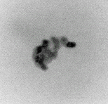
A small amount of osmium tetroxide(OsO4)gas / naphthalene gas(C10H18) is introduced into the small gas reaction vessel that equips with an anode plate and a cathode plate. Then, in the gas reaction vessel, when a DC glow discharge is generated under a thin sublimation gas pressure, the osmium metal molecules excited by the collision of electrons instantaneously become plasma between the two electrodes. The positive column and the negative glow phase are separated, and the blue-violet light of the negative glow phase emits. At the same time, the positive ion metal molecules instantly adhere to the surface of the specimen, which is placed in the negative glow phase area on the cathode plate, and an osmium metal thin film/plasma-polymerized film(naphthalene) is formed. With an osmium metal thin film on the surface of the SEM specimen, an extremely clear image can be obtained. |
![]() Application
Application
![]()
|
|
![]() The Merit of Automatic Operation
The Merit of Automatic Operation
![]()
|
Our current model of the osmium plasma coater (we have manual model in the past) is fully automatc. After placing the specimen on the sample stage, closing the gas reaction vessel and setting the thickness of the film, you can coat a film simply by pressing the start button. Since there is no complicated manual operation, there are no artificial film thickness errors. Thus, the current automatic model has excellent repeatability and higher safety.
|
|||||||||||||||||||||
|
 |
|||
|
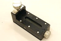
Osmium Tetroxide ampule (OsO4) |
|
|
|
|||
|
|||||||||||||
Thông số kỹ thuật chính model OPC60A:
- Buông mẫu: Làm bằng vật liệu thủy tính (glass), 120(φ) x 73(H) mm.
- Kích thước tối đa mẫu: 33(W) x 33(D) x 4(H) mm or 36(φ) x 14(H) mm.
- Gắn được các mẫu SEM kích thước: 10 mmφ x 7 pcs or 15 mmφ x 4 pcs or 36 mmφ x 1 pc.
- Bề dầy lớp phủ: Từ vài Nano tới vài trăm nm.
- Bề dầy lớp phủ có thể set nhỏ nhất: 0.1nm ở chế độ Ultra - thin film mode/ 1nm ở chế độ Normal mode.
- Loại vật liệu phủ: Màng dẫn Osmium (Osmium film).
- Đặc tính bộ OsO4: Detachable (capable to store in freezer) / Gas port integrated safety locking pin / Observation window / Built-in ampoule cutter.
- Cách đưa khí vào ra: Tự động điều khiển nhờ đầu đo chân không, solenoids và bơm hút chân không.
- Nguồn điện: 100VAC, 1 pha, 50Hz, 10A (bao gồm cả tải của bơm sơ cấp).
- Kích thước: 450(w) x 390(D) x 340(H) mm.
- Trọng lượng: 20kg.
- Thông số bơm sơ cấp: Điện áp 100VAC, 50Hz, 200W, 5.6A, tốc độ bơm 50L/min (50Hz), kích thước: 170(W) x 400(L) x 580(H) mm, trọng lượng: 18kg.
![]() Technical data
Technical data
![]()
|
OPC60A is used to coat conductive film for SEM, FE-SEM specimens. OPC80T is used to coat conductive film for SEM, FE-SEM specimens and support film for TEM specimens. If you use OPC, you can coat osmium conductive film safety and easily. You can operate OPC automatically. As long as you coat osmium of a few nm in a specimen with OPC, the specimen can get enough conductivity. Because osmium conductive film can be coated extremely uniformly. Besides osmium conductive film is no heat damage. OPC is epoch-making equipment. OPC80T can coat not only osmium conductive film but also naphthalene film. Naphthalene film is non-conductive, so strong, medicine character-resistant, and heat-resistant. Besides naphthalene film is applied as a support film. Naphthalene film is also applied as a protective film of specimens for FIB(Focused Ion Beam Equipment). ※See various technical data by clicking the following items.
|
![]() 【Publications and technical data】
【Publications and technical data】
![]()
| 1. | Samuel Angiboust & Mostafa Fayek &Ian M. Power & Alfredo Camacho & Georges Calas& Gordon Southam, Structural and biological control of the Cenozoic epithermaluranium concentrations from the Sierra Pena Blanca, MexicoMiner, Deposita December 2012, Volume 47, Issue 8, pp 859-874 | |
| 2. | Retno KAWURI, Dewa Ngurah SUPRAPTA, Youji NITTA and Takashi HOMMA, Destructive Leaf Rot Disease Caused by Fusarium oxysporum on Aloe barbadensis Miller in Bali, Agricultural Science Research Journal Vol. 2(6) pp. 295-301 June 2012 | |
| 3. | Lora L. Brehm, Towhid T. Hasan, Daniel A. Libby, Todd R. Bryden, COMPOSITION AND LAYER THICKNESS of COPPER-INDIUM-GALLIUM-SELENIUM AND MOLYBDENUM LAYERS ON GLASS BY X-RAY FLUORESCENCE SPECTROSCOPY, JCPDS-International Centre for Diffraction Data 2011 ISSN 1097-0002 | |
| 4. | Sumio Isogai, Mayuko Horiguchi, and Jiro Hitomi, The para-aortic ridge plays a key role in the formation of the renal, adrenal and gonadal vascular systems, Journal of Anatomy 2010 June; 216(6): 656-670. | |
| 5. | Chisato Takahashi, Takashi Shirai and Masayoshi Fuji, Electron Microscopic Observation of Fine Morphology of Wet Agar Gel Using a Typical Hydrophilic Ionic Liquid, 1-Butyl-3-Methylimidazolium Tetrafluoroborate, DOI 10.1007/s00126-012-0408-5 | |
| 6. | 佐藤 和彦、堀内 健、杉本 健二,硬Ⅹ線光電子分光法による高分子フィルム表面の深さ方向分析,SPring-8 重点産業利用課題成果報告書 2010A | |
| 7. | Beverly L. Falcon, Hiroya Hashizume, Petros Koumoutsakos, Jeyling Chou, James V. Bready, Angela Coxon, Jonathan D. Oliner, and Donald M. Mcdonald, Contrasting Actions of Selective Inhibitors of Angiopoietin-1 and Angiopoientin-2 on the Normalization of Tumor Blood Vessels, American Journal of Pathologu, doi : 10. 2353/ajpath. 2009. 090391 | |
| 8. | Makoto Takafuji, Naoko Azuma, Koji Miyamoto, Satoru Maeda and Hirotaka Ihara, Polycondensation and Stabilization of Chirally Ordered Molecular Organogels Derived from Alkoxysilyl Group-Containing l-Glutamide Lipid, Langmuir, Article ASAP DOI: 10.1021/la804321u Publication Date (Web) March 17 (2009) | |
| 9. | Hirotaka Ihara, Sachiyo Kubota, Atsumi Uchimura, Yuki Sakaia, Takeshi Wakiya, M. Mizanur Rahmana, Shoji Nagaokab and Makoto Takafujia, A facile preparation method for selfassembled monolayers with silica particles on polystyrene-based microspheres, Materials Chemistry and Physics Volume 114, Issue 1, Pages 1-5, 15 March (2009) | |
| 10. | E. Beach, S. Browna, K. Shqaua, M. Motterna, Z. Warchola and P. Morrisa, Solvothermal synthesis of nanostructured NiO, ZnO and Co3O4 microspheres, Materials LettersVolume 62, Issues 12-13 Pages 1957-1960, 30 April (2008) | |
| 11. | M.Murakami, Loc T. Nguyen, Zhen W. Zhang, Karen L. Moodie, P. Carmelier, Radu V. Stan and M. Simons, The FGF system has a key role in regulating vascular integrity, Journal of Investigation 118(10):3355-3366(2008) | |
| 12. | Miklos Czauna, M. Mizanur Rahmana, Makoto Takafuji and Hirotaka Ihara, Molecular shape recognition-structure correlation in a phenylalanine-based polymer_silica composite by surface-initiated atom transfer radical polymerization, Polymer Volume 49, Issue 25, Pages 5410-5416, 24 November (2008) | |
| 13. | Y. Ninomiya and S. Ichinose, Subcellular Distribution of Mitochondrial Ribosomal RNA in the Mouse Oocyte and Zygote, PLoS ONE. 2007; 2(11): e1241. | |
| 14. | Kimitake Tsuchiya, Naohiko Inase, Shizuko Ichinose, Yutaka Usui,Yasunari Miyazaki, Yoshio Ohtani, Noboru Ando, Takumi Akashi, Yasuhiro Kondoh, Hiroyuki Taniguchi and Yasuyuki Yoshizawa, Elemental analysis of inorganic dusts in lung tissues of interstitial pneumonias, J Med Dent Sci ; 54 (2007) | |
| 15. | Y. Yuan, Y. Shimada, S. Ichinose, J. Tagami, Effect of dentin depth on hybridization quality using different bonding tactics in vivo, Journal of Dentistry, Volume 35, Issue 8, Pages 664-672 (2007) | |
| 16. | P. Baluk, J. Fuxe, H. Hashizume, T. Romano, E. Lashnits, S. Butz, D. Vestweber, M. Corada, C. Molendini, E. Dejana, and Donald M. McDonald, Functionally specialized junctions between endothelial cells of lymphatic vessels, The Journal of Experimental Medicine, Vol. 204, No. 10, 2349-2362, (2007) | |
| 17. | 森 行正、阪井 博明,Osコーティングを用いたセラミック絶縁材料ナノ表面のオージェ電子分光法,セラミックデータブック2006(別刷) 工業と製品 Vol.34 No.88 工業製品技術協会(株式会社テクノプラザ),2006 Vol.34 | |
| 18. | 森 行正,Osコーティングを用いた酸化物絶縁材料・複合材料の帯電抑圧法の研究,Journal of Surface Analysis Vol. 13 No. 1 | |
| 19. | Y. Mori, Practical Technique for Restraining Differential Charging in X-ray Photoelectron Spectroscopy: Os Coating, Journal of Surface Analysis, Vol. 12 No. 2, 113-117 (2005) | |
| 20. | T. Yoshida, Ohno-Matsui,S. Ichinose, T. Sato, N. Iwata, Takaomi C. Saido,4 T. Hisatomi, M. Mochizuki, and Ikuo Morita, The potential role of amyloid β in the pathogenesis of agerelated macular degeneration, J Clin Invest. 2005 October 1; 115(10): 2793_28 | |
| 21. | Shizuko Ichinose, Kazuo Yamagata, Ichiro Sekiya, Takeshi Muneta and Motoki Tagami, Detailed examination of cartilage formation and Endochondral ossification using human mesenchymal stem cells, Clinical and Experimental Pharmacology and Physiology Volume 32 Issue 7, Pages 561 - 570 (2005) | |
| 22. | Y. Mori, S. Tanemura, Charge compensation of composite materials using Os coating in X-ray photoelectron spectroscopy, Applied Surface Science, 229, 242-248 (2004) | |
| 23. | Takahashi-Iwanaga H and Shimoda H: The three-dimensional microanatomy of Meissner corpuscles in monkey palmar skin. J Neurocytol 32: 363-371, 2003 | |
| 24. | M. Fujihira, Y. Tani, M. Furugori, U. Akiba, Y. Okabe, Chemical force microscopy of self-assembled monolayers on sputtered gold films patterned by phase separation, ultramicroscopy, 86, 63-73 (2001) | |
| 25. | 山田 豊, 中山恵美子, 後藤正和, 万木 豊, 高部圭司, 苅田修一, 藤田 稔, 担子菌類の酵素プロファイルとそのバガス分解プロセス,Grassland Science 46(3・4):265-273 (2000) | |
| 26. | Y. YAMANE, H. SHIGA, H. ASOU, H. HAGA, K. KAWABATA, K. ABE, and E. ITO, Dynamics of Astrocyte Adhesion as Analyzed by a Combination of Atomic Force Microscopy and Immuno-cytochemistry: the Involvement of Actin Filaments and Connexin 43 in the Early Stage of Adhesion, Arch. Histol. Cytol., Vol. 62, No. 4, 355-361 (1999) | |
| 27. | 岡村洋介、寺村裕治、武岡真司、土田英俊、鈴木英紀、渡辺直英、半田誠、池田康夫、フィブリノーゲンγ鎖ドデカペプチド粒子の血小板代替物の展開、人工血液 Vol.11,No.4 | |
| 28. | Hamada H, Hiroi T, Nishiyama Y, Takahashi H, Masunaga Y, Hachimura S, Kaminogawa S, Takahashi-Iwanaga H, Iwanaga T, Kiyono H, Yamamoto H and Ishikawa H: Identification of multiple isolated lymphoid follicles on the antimesenteric wall of the mouse small intestine. J Immunol 168: 57-64 |

Magnetron Sputter
Giá bán: liên hệOsmium Plasma Coater
Giá bán: liên hệ
- Tin Tức Nổi Bật

AMETEK VIETNAM
26/10/2024
EDAX Vietnam
26/10/2024
Túi khí chèn hàng container
21/11/2022
Đá mài kim cương là gì
15/11/2022

